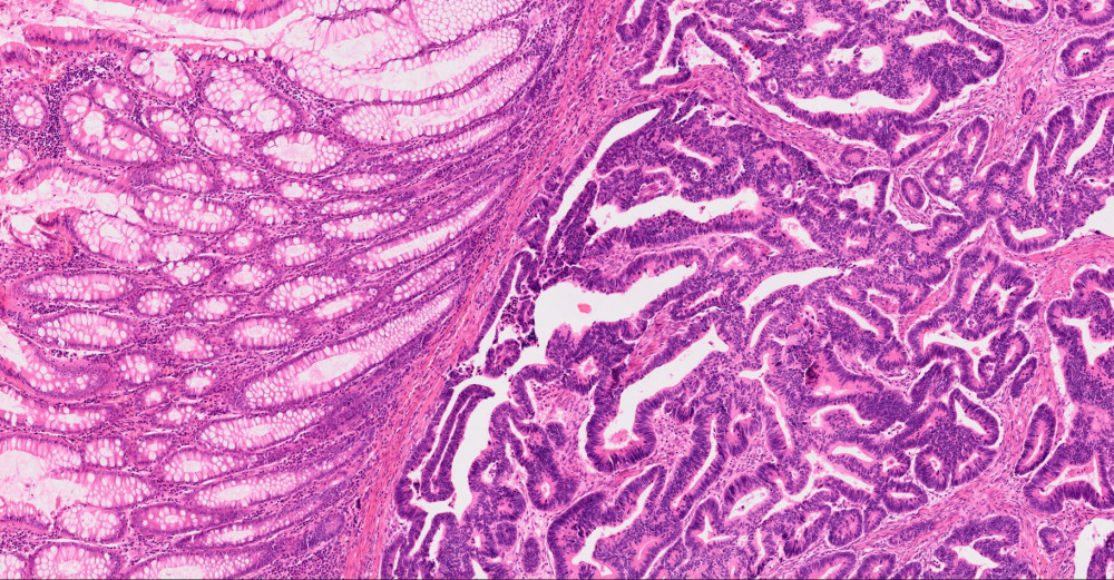
In March, we are presenting a photograph taken by Esraa Ali, a doctoral student from the Translational Cancer Genomics Lab. This light microscopy image shows an example of the difference between the normal mucosa of the colon (on the left) and colon cancer (on the right). The normal mucosa is organized with stereotypical crypts that contain several thousands of cells arranged in the form of a test-tube open to the lumen of the colon. In the tumor, they become distorted, irregular, stained darkly, and show abnormality at the cellular level.